1800 867 1390
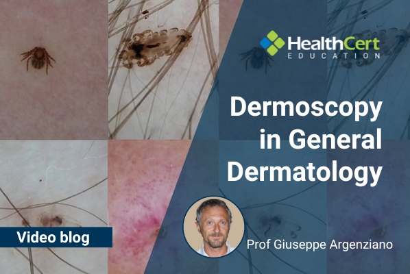
Dermoscopy update: Prof Giuseppe Argenziano explores further general dermatology conditions diagnosed through dermoscopy, including alopecia.
HealthCert Education
Tune into this latest dermoscopy update video in which Professor Giuseppe Argenziano continues last month's exploration of general dermatology conditions that can be diagnosed through the use of dermoscopy, including pigmented purpuric dermatitis, porokeratosis, sarcoidosis, alopecia, and more.
Learn more about this topic in the HealthCert Professional Diploma program in Dermoscopy, providing tailored medical dermoscopy training online for GPs.
In the video, Prof Argenziano explores pigmented purpuric dermatitis, which includes:
- Lichen aureus
- Lichenoid purpura Gougerot-Blum
- Eczematid-like purpur
- Majocchi and Schamberg diseases
These lesions are characterised by two clues: brown pigmentation and purpuric dots which don't go away when you press with your dermatoscope. This skin condition is a spring and summertime disease often related to sweating. It can cause itching and is often found on the trunk.
He also covers porokeratosis (which is characterised by a ring-like scale) and cutaneous sarcoidosis (indicated by an orange colour and sharp vessels).
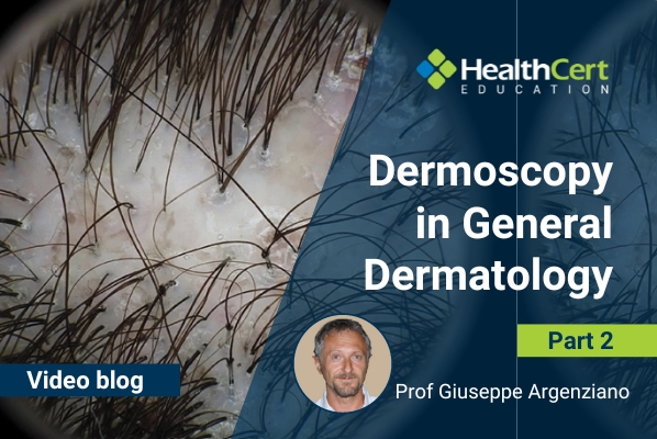
Hair conditions are also covered, including alopecia areata (characterised by hair abnormalities and yellow dots). Prof Argenziano explains how dermoscopy in these cases can actually change your opinion of the diagnosis by adding more information and diving deeper than superficial clinical examination.
Prof Argenziano takes us through some real patient cases and highlights the dermoscopic clues which indicate these general dermatology conditions to aid with primary care diagnosis.
See all this and much more in the full video below!
Watch the full video now:
For further information on this topic, you may be interested to learn more about the HealthCert Professional Diploma program in Dermoscopy, providing tailored medical dermoscopy training online for GPs.
 Prof Giuseppe Argenziano is Professor and Head of the Dermatology Unit at the University of Campania, Naples, Italy; Co-founder and past president of the International Dermoscopy Society; and Editor-in-Chief of Dermatology Practical and Conceptual Journal. His main research field is dermato-oncology, authoring numerous scientific articles and books concerning dermoscopy, melanoma and non-melanoma skin cancer. As coordinator of the Melanoma Unit at the Campania University, he has established a successful tertiary, multidisciplinary, referral center particularly devoted to the diagnosis and management of patients with melanoma and non-melanoma skin cancer.
Over the past 25 years, Prof Argenziano has supervised over 500 foreign and Italian residents in dermatology, established scientific collaborations with 1500+ colleagues from more than 50 nations, and organised more than 500 national and international didactic meetings, courses and conferences (such as the Consensus Net Meeting on Dermoscopy and the First Congress of the International Dermoscopy Society).
Prof Argenziano has authored more than 650 full scientific articles and produced landmark primary publications and books in the field of melanoma and dermoscopy. Over the past 25 years he has been invited as speaker and/or chairman in more than 500 national and international conferences in the field of dermatology. His combined publications have received a sum total of 15.250+ citations with an h-index value of 61 (Scopus 2020).