This month we further explore the topic of Total Body Photography (TBP) and its role in skin cancer (especially melanoma) surveillance, identification and diagnosis. The topic is important and rapidly developing, and it has supporters on all sides.
For more information on this topic, you may be interested in the HealthCert Professional Diploma in Skin Cancer Medicine: medical courses online + with optional workshops ideal for GP training towards a focused expertise in skin cancer.
It seems obvious that taking photographs and having software or artificial intelligence run over the images must be a good thing. Studies have been done, of course, and there is some compelling data. However, definitive data is lacking because controlled studies are lacking. Simply put, the ideal study would compare normal care against normal care with TBP.
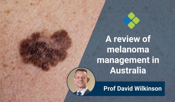
This latest paper from Brown et al in the Australasian Journal of Dermatology is quite intriguing. Data from a dermatology practice expert in Brisbane provides data that shows a much higher number of in situ melanomas (and ratio to invasive) than those reported in TBP studies.
It is also instructive to read that about half of all melanomas (in situ and invasive) were diagnosed through shave biopsy usually done at the time of the consultation.
A fascinating paper reporting on real-world, real-life practice. Worth a careful read and reflection.
Read the full paper here.
– Prof David Wilkinson
For further information on this topic, you may be interested in the HealthCert Professional Diploma in Skin Cancer Medicine.


 1800 867 1390

