
 1800 867 1390
1 minute read
Skin & Skin Cancer Conference with Prof Cliff Rosendahl
Prof Cliff Rosendahl invites you to join him at HealthCert's Skin & Skin Cancer Conference upcoming in Brisbane in July 2024.
Prof Cliff Rosendahl invites you to join him at HealthCert's Skin & Skin Cancer Conference upcoming in Brisbane in July 2024.
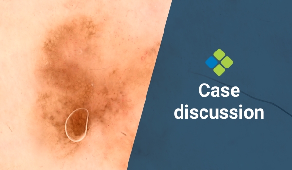
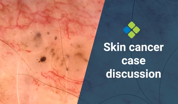
Case discussion: 5x4mm flat pigmented lesion found on posterior right leg of 65-year-old female. What do you think?
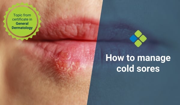
Read a simple GP guide to manage cold sores, including how to recognise symptoms, establish a diagnosis, educate patients, provide treatment and follow-up.

Dr Joe Kosterich talks about medical cannabis: myths and misconceptions, how it's used as medicine, and how doctors in Australia can start prescribing it.
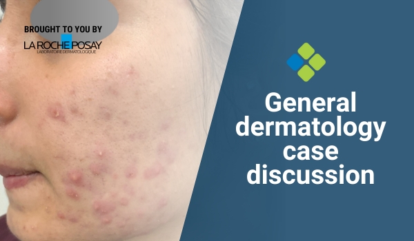
Case discussion: A 29-year-old female patient of Indian heritage presents with moderate papulopustular acne. What is your management plan?
Case discussion: A 4x3mm lesion is identified on the clavicle of a 40-year-old male during a routine skin check. What do you think of it?